The Single Strategy To Use For Spectrophotometers
The Single Strategy To Use For Spectrophotometers
Blog Article
What Does Spectrophotometers Do?
Table of ContentsAn Unbiased View of Uv/visGet This Report on Circular DichroismThe smart Trick of Circularly Polarized Luminescence That Nobody is DiscussingUv/vis - An OverviewCircularly Polarized Luminescence Things To Know Before You BuyUv/vis/nir Things To Know Before You BuyFacts About Uv/vis UncoveredCircularly Polarized Luminescence for DummiesThe 2-Minute Rule for Circular Dichroism7 Simple Techniques For Uv/vis/nirA Biased View of Circular DichroismEverything about Circular DichroismUv/vis Fundamentals Explained
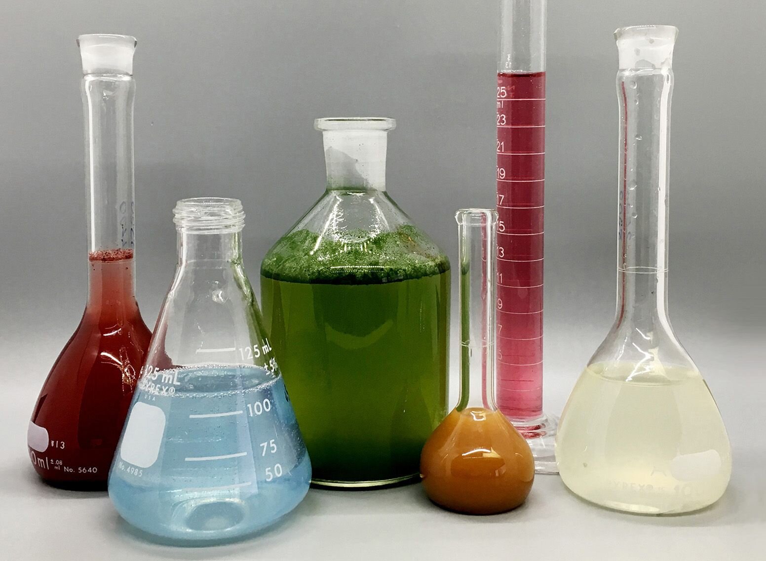
It is then scanned through the sample and the recommendation services. Fractions of the occurrence wavelengths are sent through, or reflected from, the sample and the referral. Electronic circuits convert the relative currents into direct transmission percentages and/or absorbance/concentration values.The transmission of a referral compound is set as a standard (datum) worth, so the transmission of all other substances are tape-recorded relative to the initial "zeroed" compound. The spectrophotometer then converts the transmission ratio into 'absorbency', the concentration of specific parts of the test sample relative to the initial substance.
Because samples in these applications are not easily available in large amounts, they are particularly matched to being evaluated in this non-destructive strategy. In addition, precious sample can be saved by making use of a micro-volume platform where as low as 1u, L of sample is required for total analyses. A brief description of the treatment of spectrophotometry includes comparing the absorbency of a blank sample that does not consist of a colored substance to a sample which contains a colored compound.
Indicators on Circular Dichroism You Should Know
In biochemical experiments, a chemical and/or physical residential or commercial property is chosen and the treatment that is utilized is specific to that residential or commercial property in order to derive more information about the sample, such as the amount, pureness, enzyme activity, etc. Spectrophotometry can be utilized for a number of strategies such as identifying optimal wavelength absorbance of samples, identifying optimal p, H for absorbance of samples, figuring out concentrations of unknown samples, and identifying the p, Ka of different samples.: 21119 Spectrophotometry is likewise a practical procedure for protein purification and can also be utilized as a technique to develop optical assays of a compound.
It is possible to understand the concentrations of a two component mixture utilizing the absorption spectra of the basic solutions of each part. To do this, it is essential to understand the extinction coefficient of this mixture at 2 wave lengths and the extinction coefficients of options that include the recognized weights of the two components.
The 4-Minute Rule for Circular Dichroism
Most spectrophotometers are used in the UV and noticeable areas of the spectrum, and some of these instruments likewise run into the near-infrared Region. The concentration of a protein can be estimated by determining the OD at 280 nm due to the presence of tryptophan, tyrosine and phenylalanine (https://hubpages.com/@olisclarity1).
This method needs a spectrophotometer capable of measuring in the UV area with quartz cuvettes.: 135 Ultraviolet-visible (UV-vis) spectroscopy involves energy levels that excite electronic shifts. Absorption of UV-vis light delights molecules that are in ground-states to their excited-states.
20. 8 O.D. Ink manufacturers, printing business, textiles suppliers, and numerous more, require the data offered through colorimetry. They take readings in the area of every 520 nanometers along the noticeable region, and produce a spectral reflectance curve or an information stream for alternative presentations. These curves can be utilized to test a brand-new batch of colorant to inspect if it makes a match to requirements, e.
3 Simple Techniques For Uv/vis
Traditional noticeable area spectrophotometers can not find if a colorant or the base material has fluorescence. This can make it difficult to handle color concerns if for example several of the printing inks is fluorescent. Where a colorant contains fluorescence, a bi-spectral fluorescent spectrophotometer is utilized (https://www.slideshare.net/julieanndesalorenz30). There are 2 significant setups for visual spectrum spectrophotometers, d/8 (spherical) and 0/45.
Scientists utilize this instrument to determine the quantity of compounds in a sample. If the compound is more concentrated more light will be taken in by the sample; within little varieties, the Beer, Lambert law holds and the absorbance in between samples differ with concentration linearly. In the case of printing measurements two alternative settings are commonly utilized- without/with uv filter to manage better the effect of uv brighteners within the paper stock.
The Spectrophotometers Statements
Some applications need little volume measurements which can be carried out with micro-volume platforms. As described in the applications area, spectrophotometry can be used in both qualitative and quantitative analysis of DNA, RNA, and proteins. Qualitative analysis can be used and spectrophotometers are used to tape-record spectra of compounds by scanning broad wavelength regions to identify the absorbance residential or commercial properties (the strength of the color) of the compound at each wavelength.

Top Guidelines Of Uv/vis
One significant factor is the type of photosensors that are offered for various spectral areas, however infrared measurement is also difficult because virtually whatever gives off IR as thermal radiation, particularly at wavelengths beyond about 5 m. Another problem is that many materials such as glass and plastic take in infrared, making it incompatible as an optical medium.
Recovered Dec 23, 2018. Essential Lab Techniques for Biochemistry and Biotechnology (Second ed.). The vital guide to analytical chemistry.
Oke, J. B.; Gunn, J. E.
3 Simple Techniques For Uv/vis/nir
1021/ac50048a728. ISSN0003-2700. Ninfa AJ, Ballou DP, Benore M (2015 ). Fundamental Laboratory Methods for Biochemistry and Biotechnology (3, rev. ed.). Hoboken, NJ: Wiley & Sons. p. 77. ISBN9780470924525. OCLC915641828. "Completely Automatic Double Beam - Atomic Absorption Spectrophotometer (AA 8000)". Lab Devices. Labindia Analytical Instruments Pvt. Ltd. "Spectrophotometry Applications and Principles".
The Definitive Guide to Circular Dichroism
Obtained Jul 4, 2018. Trumbo, Toni A.; Schultz, Emeric; Borland, Michael G.; Pugh, Michael Eugene (April 27, 2013). "Applied Spectrophotometry: Analysis of a Biochemical Mix". Biochemistry and Molecular Biology Education. 41 (4 ): 24250. doi:10. 1002/bmb. 20694. PMID 23625877. (PDF). www. mt.com. Mettler-Toledo AG, Analytical. 2016. Obtained Dec 23, 2018. Cortez, C.; Szepaniuk, A.; Gomes da Silva, L.
"Checking Out Proteins Filtration Strategies Animations as Tools for the Biochemistry Mentor". Journal of Biochemistry Education. 8 (2 ): 12. doi:. Garrett RH, Grisham CM (2013 ). Biochemistry. Belmont, CA: Cengage. p. 106. ISBN 978-1133106296. OCLC 801650341. Vacation, Ensor Roslyn (May 27, 1936). "Spectrophotometry of proteins". Biochemical Journal. 30 (10 ): 17951803. doi:10. 1042/bj0301795.
PMID 16746224. Hermannsson, Ptur G.; Vannahme, Christoph; Smith, Cameron L. C.; Srensen, Kristian T.; Kristensen, Anders (2015 ). "Refractive index dispersion picking up utilizing a selection of photonic crystal resonant reflectors". Applied Physics Letters. 107 (6 ): 061101. Bibcode:2015 Ap, Ph, L. 107f1101H. doi:10. 1063/1. 4928548. S2CID 62897708. Mavrodineanu R, Schultz JI, Menis O, eds.
Indicators on Circularly Polarized Luminescence You Should Know
U.S. Department of Commerce National Bureau of Standards unique publication; 378. Washington, D.C.: U.S. National Bureau of Standards.
The procedure starts with a regulated source of light that brightens the examined sample. When it comes to reflection, as this light connects with the sample, some is absorbed or given off. The given off light travels to the detector, which is examined, measured, and provided as industry-standard color scales and indices.
Industry governing bodies generally define specific metrics for particular products, such as Tomato and Coffee indices. The streamlined math appears like this: Where R is the reflection coefficient. All terms are evaluated over the noticeable spectrum from 400 to 700 nm. When it comes to transmission, when the light interacts with the sample, it is either absorbed, reflected, or sent.
Everything about Circularly Polarized Luminescence
Examples include APHA (American Public Health Association) for watercolor and pureness analysis, ASTM D1500 for petrochemical color analysis, edible oil indices utilized in food, and color analyses of drinks. The simplified math looks like this:. Where T is the transmission coefficient. All terms are assessed over the visible spectrum from 400 to 700 nm.
Image Credit: Matej Kastelic/ Dr. Arnold J. Beckman and his associates at the National Technologies Laboratories first developed the spectrophotometer in 1940. In 1935 Beckman established the business, and the discovery of the spectrophotometer was their most ground-breaking development.
Not known Facts About Uv/vis
Over time, researchers kept improving the spectrophotometer style to improve its efficiency. The UV capabilities of the design B spectrophotometer were enhanced by replacing the glass prism with a quartz prism.
Normally, a spectrophotometer is made up of two instruments, particularly, a spectrometer and a photometer. A fundamental spectrophotometer consists of a light source, a monochromator, a collimator for dig this straight light beam transmission, a cuvette to place a sample, and a photoelectric detector.
Uv/vis/nir Can Be Fun For Anyone
There are various kinds of spectrophotometers in various sizes and shapes, each with its own purpose or performance. A spectrophotometer determines just how much light is shown by chemical elements. spectrophotometers. It determines the difference in light strength based on the overall quantity of light presented to a sample and the amount of beam that passes through the sample solution
As per the instrument's design, the sample is placed between the spectrometer and the photometer. After the light is passed through the sample, the photometer measures its strength and shows the reading. A spectrophotometer is used to figure out the concentration of both colorless and colored solutes in a solution. This instrument is used to identify the rate of a response.
Report this page